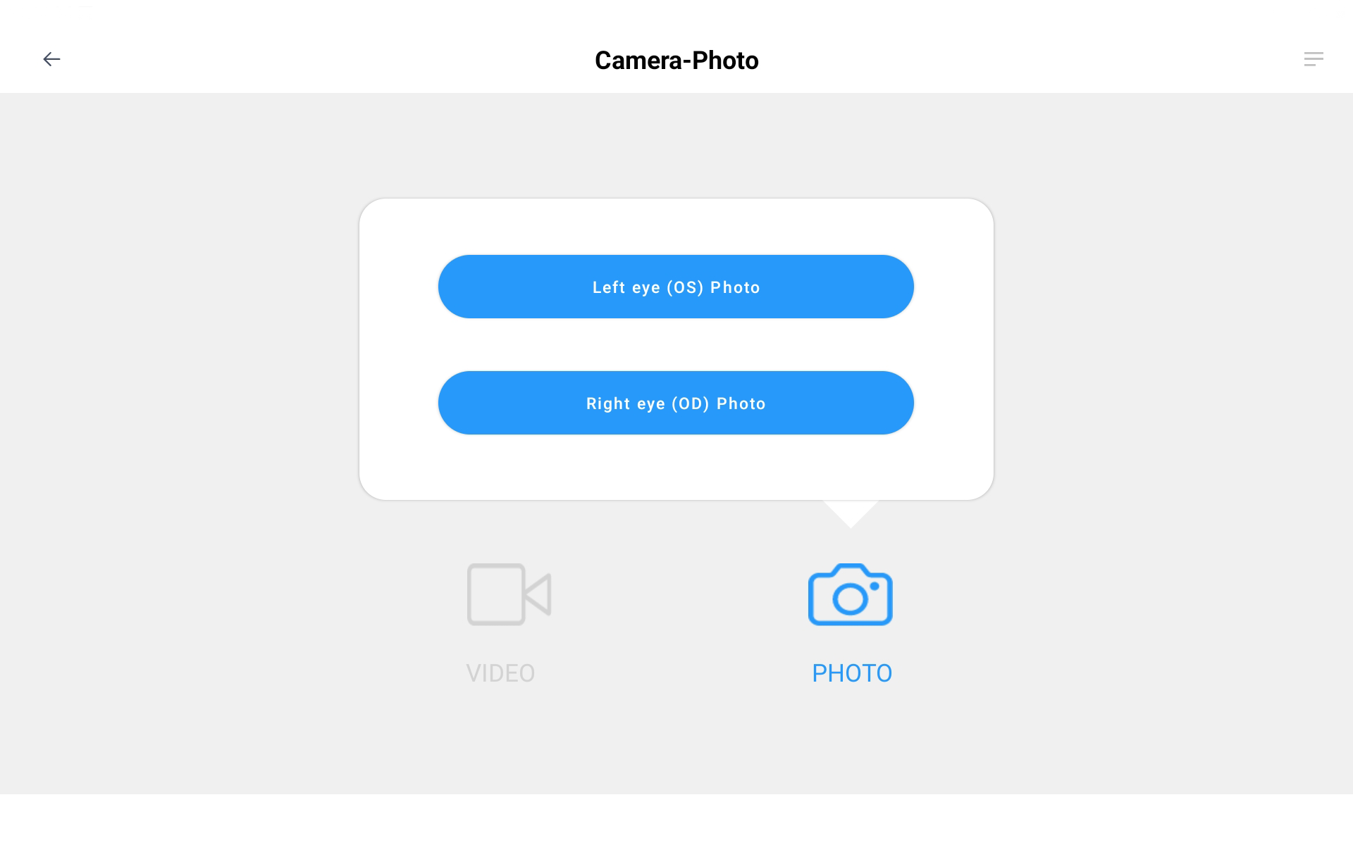
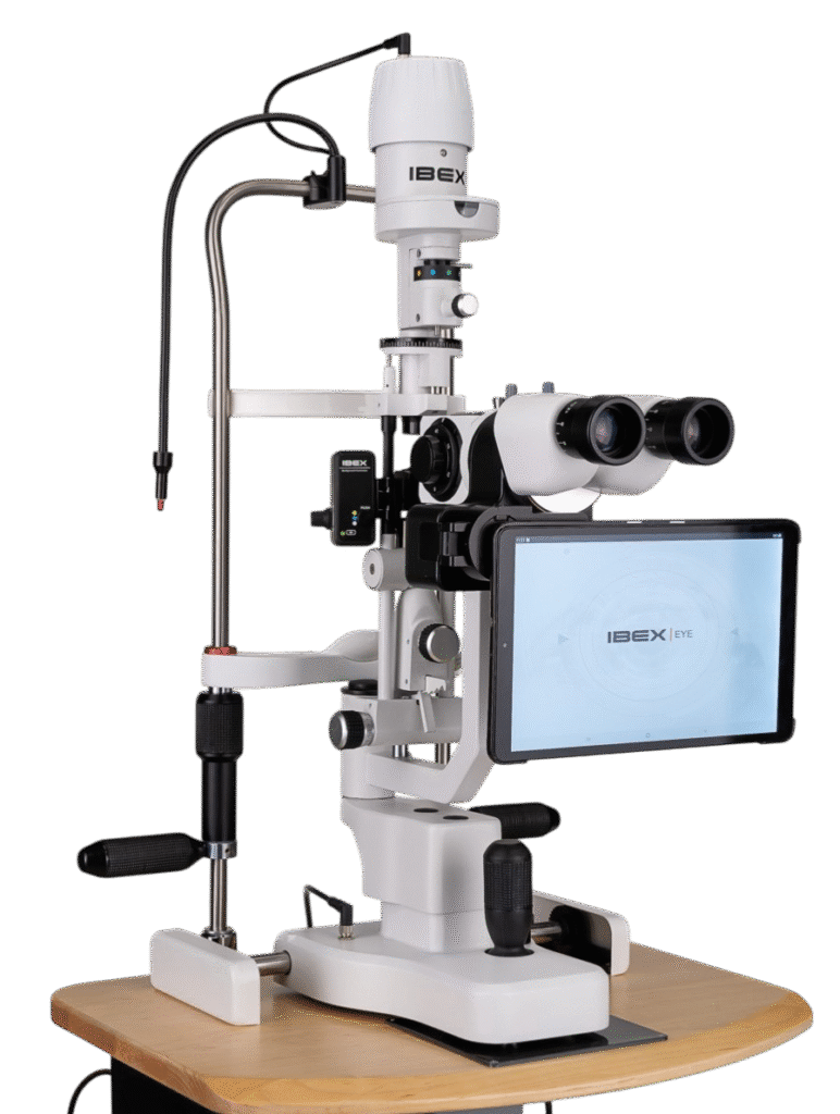
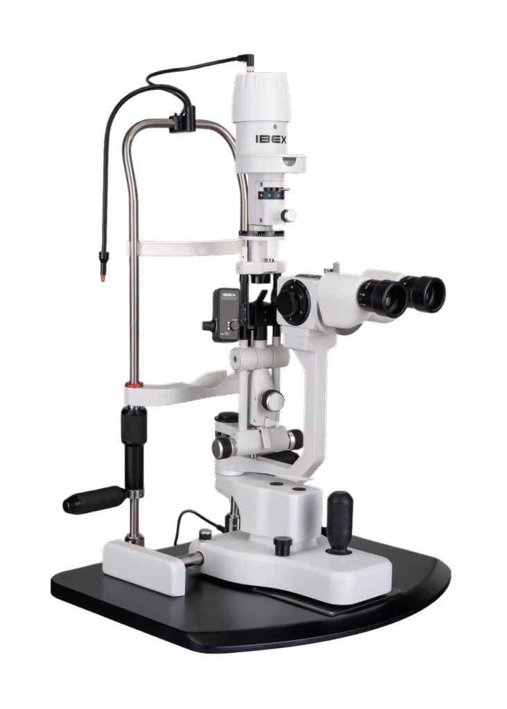
Opteon™ Slit Lamp Imaging + Meibography

Photos & Videos. Clinical Documentation. Patient Education. Reimbursement.

Illuminate your exams and transform patient engagement like never before. When patients see detailed images of their eyes, they gain a deeper understanding of their health and recognize the importance of compliance and follow-up care. Throughout development, our team saw firsthand the profound impact of imaging in the clinic—watching patients smile, express genuine appreciation, and embrace their health journey. That unforgettable experience remains the most rewarding part of our work.

Opteon was born and developed in the clinic, meticulously tested, and perfected through countless hours to ensure that it flows with your exam routine — delivering unmatched reliability and efficiency.


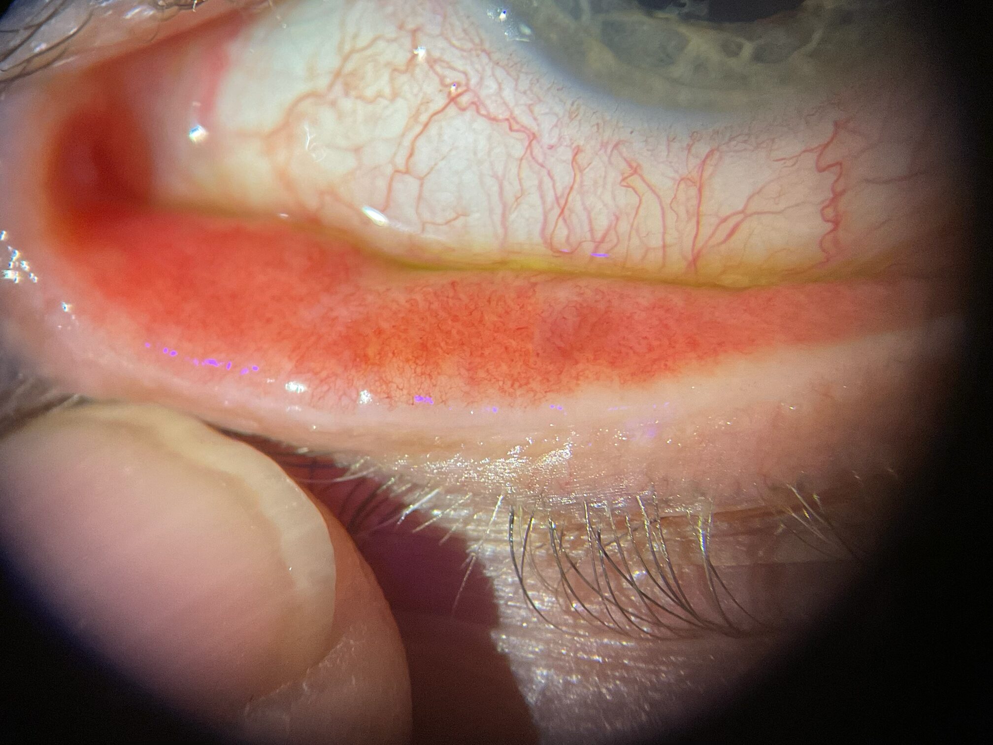
Pushing ahead, our imaging system includes our new, patent-pending Halo background light. Working with native slit lamp illumination, Halo delivers unparalleled clarity of the ocular adnexa. You’ll uncover details of the meibomian glands, acinar spaces, conjunctiva, lid margins, and lashes like never before.

Potential Reimbursement:

CPT / HCPCS Code 92285 External ocular photography with interpretation and report for documentation of medical progress, (eg close-up photography, slit lamp photography, goniophotography, stereo photography).

Click to View – Billing and Coding: Ocular Photography – External (A57068) for a complete listing of codes that
support medical necessity.

Opteon imaging includes our revolutionary Halo™ Background Light

Providing wide-field views of the ocular adnexa, you’ll witness morphology
of the meibomian glands in vivo, and truly exceptional tear film patterns

The eye lids are essential to ocular health, yet too often receive insufficient attention during routine eye exams. They serve not only as protective barriers for the ocular surface, but also as early indicators of local and systemic conditions, including Dry Eye.

Halo’s light seamlessly blends with native slit lamp illumination, delivering unparalleled clarity in the ocular adnexa. With diffused, full-field lighting, you’ll uncover details of the meibomian glands, acinar spaces, conjunctiva, lid margins, and lashes like never before.

Through a comparative study of established infrared (IR) photography and Halo’s red-free meibography, we identified remarkable advantages. While IR meibography visualizes central gland tubes, Halo goes further and illuminates the acinar spaces — critical structures responsible for meibum production and potentially offering greater diagnostic insight.

Plus, in a single touch, our imaging software converts color images to red-free, black and white images. A second touch inverts the black and white image, revealing vessels and meibomian blood supply for the first time, a vital factor in gland function. This technique unveils numerous accessory lacrimal glands, offering deeper perspectives into ocular surface disorders.

Halo fits the vast majority of slit lamps. Patent-Pending.

Potential Reimbursement:

CPT / HCPCS Code 92285 External ocular photography with interpretation and report for documentation of medical
progress, (eg close-up photography, slit lamp photography, goniophotography, stereo photography). including but not limited to meibography, vascular abnormalities, dry eye syndrome, lacrimal gland atrophy, conjunctivitis, blepharitis. bulbar and palpebral conjunctiva (eye redness), eyelashes (Demodex), corneal staining (fluorescein), and tear meniscus.

Click to View – Billing and Coding: Ocular Photography – External (A57068) for a complete listing of codes that support medical necessity.

You can take professional grade photos and educate patients in seconds.

A comprehensive educational image library at your fingertips

We included an extensive library with 129 high-quality clinical photos, designed to enhance diagnostics and patient education. Whether illustrating clinical findings, guiding through operative steps, or showcasing postoperative results, these images provide invaluable visual clarity—helping clinicians and patients alike understand every stage of care.

Video record and photograph tear film

Millions suffer from dry eye symptoms—burning, itching, blurred vision, and that persistent gritty sensation. Halo’s blue light highlights tear film and empowers clinicians to assess tear production, distribution, clearance, superficial punctate staining, micropsphericals, and more with accuracy and speed.

Tears not only provide eye comfort, they promote wound healing, suppress inflammation, scavenge free radicals, and defend against microbial infection.¹

Biological Functions of Tear Film, Stephen C Pflugfelder , Michael E Stern


Unveiling hidden ocular disorders

In a single touch, our imaging software converts color images to red-free, black and white images. A second touch inverts the black and white image, revealing vessels and meibomian blood supply for the first time, a vital factor in gland function. This technique unveils numerous accessory lacrimal glands, offering deeper perspectives into ocular disorders

Born in the Clinic™ software that’s intuitive and efficient

With an intuitive interface, capturing high-quality photos and videos is amazingly simple. From the moment you first turn it on, it’s intuitive, fast, and fun for all clinicians.

Ready when you are, our software rests in sleep mode, on stand-by, then, with a single touch, you’re taking high-quality photos and videos – and providing amazing patient education.

See 2 minute demonstration video on the right on how easy it is to take photos, manage, send and move to the patient EMR record.

Efficiently transfer images into your EMR

Samsung Flow wirelessly transfers images from the tablet to a designated folder on your PC or laptop. Thereafter, at your PC,
using simple drag-and-drop functionality or direct uploading, you and your team can move images seamlessly into your EMR—
streamlining documentation and eliminating hassle.

Video record, bookmark and save – you’ll never miss a shot again

While video recording your exams, bookmark and save key photo frames instantly using either the Bluetooth footswitch or the
tablet’s touchscreen. Each saved frame is marked directly on the video and moved to the screen for immediate review,
ensuring you always get the perfect shot—when it matters most.

Create a dynamic exam room and live stream directly to a large-screen Smart TV

Ideal for patient education, boosting family engagement, and enhancing professional collaboration, this seamless integration
empowers you to share insights, clarify diagnoses, and deliver clear, evidence-based treatment protocol recommendations

A remarkable camera within an all-in-one tablet

The tablet’s camera and its advanced algorithms enable you to instantly capture detailed, clear photos and videos.

For good reasons, today’s modern clinics and hospitals rely on powerful tablets with touchscreen technology to streamline
workflows and enhance efficiency: They’re beautiful, intuitive, packed with features, affordable, upgradable and replaceable.
Their all-in-one design seamlessly integrates a robust camera, operating system, and crystal clear display—eliminating the
need for external wires, computers, and monitors.

A beam splitter that puts you in control

The beam splitter is a vital element in slit lamp imaging. Our specialized, dual-purpose design ensures a standard, 100%
unfiltered view of your patient’s eyes through your slit lamp’s optics. When you’re ready to capture an image, simply slide the
selector knob to share your view with the camera—giving you complete control over your viewing experience.

Optimize space effortlessly

With Opteon, the tablet is recessed beneath the eyepieces so that clinicians can maintain comfortable, traditional viewing
while having advanced imaging at their fingertips. The display provides a generous view for both clinicians and patient
engagement, yet remains compact enough to ensure effortless access to the eyes— it’s comfort without compromise.

Compatible with many brands of parallel optic slit lamps in the market.

Click to see if your Opteon fits your lamp.

We made the set-up simple. Go from box to taking photos in less than 60 Seconds.

Click here to view the installation video

Opteon™ Premium Slit Lamp Imaging for Ocular Photography

Opteon intelligently positions the tablet below the eyepieces and recessed, where it rests
quietly and on-call until needed. This specialized design enables seamless observation through the eyepieces, or with the large screen tablet. It’s a wonderful balance of traditional eyepiece functionality with modern digital engagement.

Opteon Premium Imaging Kit includes the following components:

Halo background light
Beam splitter with 100% direct view or 60/40 share with the camera
10.5” Samsung touch-screen tablet with IBEXeye Video Cam Software
Samsung Flow App moves photos from the tablet to PC/Desktop, then to EMR
Bluetooth foot switch for hands-free start/stop video recording or taking photos

Opteon works seamlessly with various brands of parallel optic slit lamps, including our IBEX lamps, as well as models by Topcon, Haag-Streit, Marco and more. Click to see if your slit lamp is compatible.

Opteon™ Premium Slit Lamp Imaging Kit – Only $6,995.00

Or Build a Quote

Please see below for our Opteon Premium complete slit lamp imaging packages

Opteon™ Premium Slit Lamp Imaging Kit – $6,995.00
IBEX 5-Step Tower Slit Lamp – $6,995.00
Your Choice of Natural Wood or Onyx Slit Lamp Base
Only $13,990.00
Or Build a Quote
Opteon™ Premium Slit Lamp Imaging Kit – $6,995.00
IBEX 5-Step Compact Slit Lamp – $5,995.00
Your Choice of Natural Wood or Onyx Slit Lamp Base
Only $12,990.00
Or Build a Quote

Opteon™ On-Demand Slit Lamp Imaging for Ocular Photography Designed specifically for our IBEX 2-Step Tower and Compact Slit Lamps

You simply place the tablet over the slit lamp eyepieces when you wish to capture important photos or videos— it’s temporary, manual on/off, but we still includes all of Opteon’s amazing features and capabilities. It’s a terrific way to get started with slit lamp photography.

Opteon On-Demand Imaging Kit includes the following components:

Halo background light
Eyepiece mount
10.5” Samsung touch-screen tablet with IBEXeye Video Cam Software APP
Samsung Flow App moves photos from the tablet to PC/Desktop, then to EMR
Bluetooth foot switch for hands-free start/stop video recording or taking photos

Opteon™ On-Demand Slit Lamp Imaging – Only $4,495.00

Or Build a Quote

Please see below for our Opteon Premium complete slit lamp imaging packages

Opteon™ On-Demand Slit Lamp Imaging – $4,495.00
IBEX 2-Step Tower Slit Lamp – $4,995.00
Your Choice of Natural Wood or Onyx Slit Lamp Base
Only $9,490.00
Or Build a Quote
Opteon™ On-Demand Slit Lamp Imaging – $4,495.00
IBEX 2-Step Compact Slit Lamp – $3,995.00
Your Choice of Natural Wood or Onyx Slit Lamp Base
Only $8,490.00
Or Build a Quote